
Why a torn Achilles may heal so slowly
Collaborators

Research by the Wu Tsai Human Performance Alliance points to the lymphatic system as an overlooked factor in why Achilles tendon injuries heal so poorly.
By Kristy Hamilton
Canva pro images
The Achilles tendon is an anatomical marvel and, too often, a source of misery. It is the largest tendon in the human body and one of the most frequently injured. A year after a rupture, patients still report scoring around 18 percent below a full recovery, and that score doesn’t tend to improve much with time.
The standard explanation was blood supply. The Achilles doesn’t get much of it, the thinking went, so it’s slow to heal. But a study conducted in rats from the University of Oregon, published in the Journal of Orthopaedic Research, suggests it’s more complex: the problem may also lie in the lymphatic fluid that fails to drain out of the tissue.
“When tissue is injured, it produces a constant stream of waste — tissue debris, dead cells, and other byproducts — that must be removed,” said Nick Willett, Associate Director of the Wu Tsai Human Performance Alliance at Oregon and Associate Professor of Bioengineering. “When that fluid clearance is impaired, waste can’t leave the environment.” Neither can the inflammatory signals in that fluid, keeping the tissue in an inflamed state instead of moving on to repair.
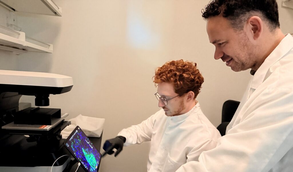
Nick Willett, an Associate Professor of Bioengineering at the University of Oregon and Associate Director of the Wu Tsai Human Performance Alliance at Oregon, and graduate student Jarod Forer examine cross-sectional images of the Achilles tendon. Courtesy University of Oregon
Tracking the fluid with glowing dye
To track both blood and lymphatic drainage, the team injected two dyes that glow under near-infrared light into the rats’ Achilles tendons. One dye was a small molecule, the other large. That size difference sends them on two different routes. The small molecule drains mainly through blood vessels (the venous route), clearing in about 1.5 hours in healthy tendons, while the larger molecule must wait for the slower lymphatic vessels, taking around 10 hours. Injecting both at once let the team watch the routes side by side.
Initially, the lymphatic dye cleared faster in the ruptured tendons than in the healthy ones. This was expected based on previous experimental findings that damaged tissue sprouts new lymphatic vessels and its disrupted outer sheath gets leakier.
Yet surprisingly, at eight weeks, instead of dropping back down to the clearance levels of the healthy controls, lymphatic drainage slowed below that of healthy controls. The tissue stayed swollen and pale. Whatever healing had occurred, the tendon still couldn’t manage its own fluid.
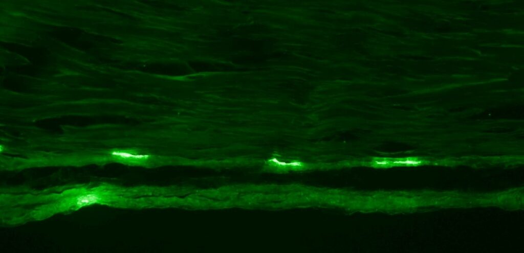
A cross-sectional image of a tendon. The cells that express the gene Prox1, such as lymphatic endothelial cells, glow green. Note that the bright green cells are localized to the tendon periphery or sheath. Courtesy Jarod Forer
Looking healed isn’t the same as being healed
The team also tracked the more familiar signs of recovery. Weight-bearing returned to normal within four weeks. Pain sensitivity and stiffness recovered by eight. But two things didn’t bounce back: the tendon’s underlying material strength and its ability to drain fluid. The results suggest that a patient can look fully recovered by one measure while remaining impaired by another.
There were also expected results involving the healthy leg: its venous (blood) drainage slowed at four and eight weeks, likely because the rats put extra load on their good leg while the hurt one healed, possibly disturbing its circulation. It’s a reminder that an injury on one side of the body doesn’t always stay on that side.
A possible target for treatment
Why should trapped fluid stall healing at all? It comes down to timing.
“Healing depends on tight coordination between an initial inflammatory response and a transition to a regenerative one,” said Willett. “If fluid clearance is impaired, those inflammatory cues persist, driving chronic inflammation that stalls the regenerative response. This is precisely what happens in tendinopathy.”
The findings come with limitations. The rats’ injured legs were never put in casts like human patients usually are, which can alter how tendons heal. The rats were followed for only eight weeks, so what happens to drainage after that is unknown.
Even so, the study put a familiar problem in a new light. If poor drainage is part of why tendons heal slowly, then the trapped fluid becomes something to treat. The researchers suspect getting fluid moving again, perhaps through the right rehabilitation exercises, might speed recovery.
“We are now working on these exact questions and beginning to investigate these findings in human subjects,” said Willett. “We are particularly interested in rehabilitative, exercise-based approaches and whether they can drive fluid clearance in both rodent and human tendon.”
Study authors include Jarod Forer, Kaitlyn Link, Bella Yannello, and Michael Hahn. This research is part of the Wu Tsai Human Performance Alliance’s Regenerative Rehabilitation moonshot.
Latest News

September 24, 2026
Wu Tsai Human Performance Alliance welcomes Emory Human Performance Institute, expanding Innovation Hub network

September 22, 2026
Alliance Round-Up: REDs risk markers, heat and iron absorption, and more

September 8, 2026
A wearable device tracked over 42,000 menstrual cycles. Here’s what researchers found
Get Engaged
Join our mailing list to receive the latest information and updates on the Wu Tsai Human Performance Alliance.